Figure 5.4 · Thesis 4 · Orayech 2015
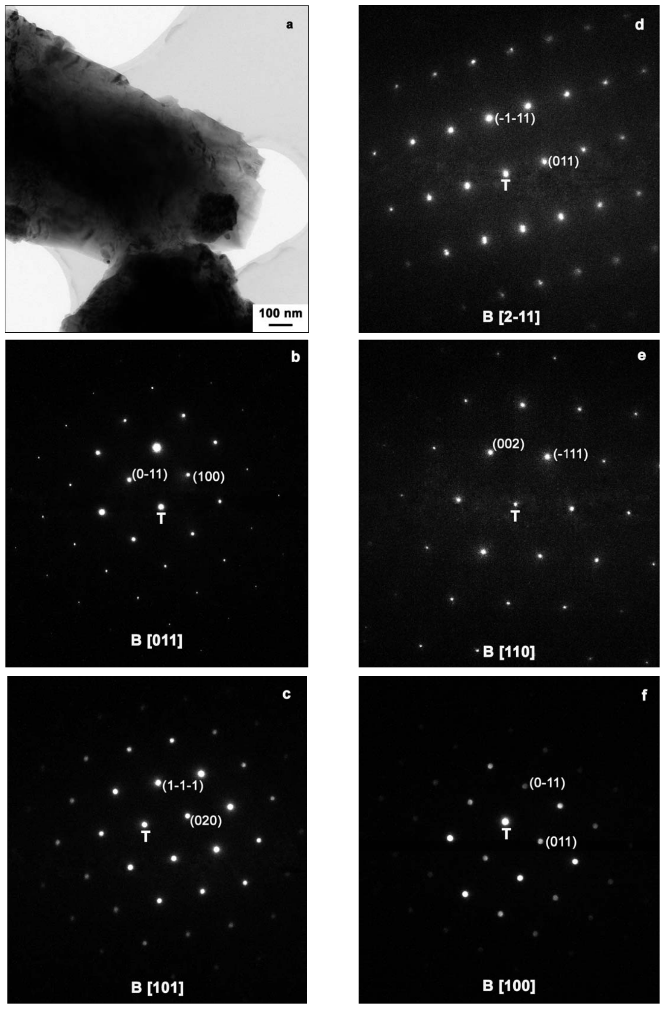
a) Bright field TEM micrograph of a typical particle in the sample. b), d) and e) SAD patterns acquired along the [011], [2-11] and [110] zone axes, respectively. c) and f) electron microdiffraction acquired along the [101] and [100] zone axes, respectively. The reflections indicated in the patterns were identified by simulations done with the JEMS software, using the neutron data presented in the previous sections.
- Document
- Thesis 4 · Orayech 2015
- Figure
- 5.4
- Kind
- tem
- Rights
- own
- Reproduce
- not-needed
auto-cropped from the OCR Markdown crop box (stage B); materials and kind to be reviewed in Phase 1
Evidence
- doc:thesis-04 · figure 5.4 · page 82 · OCR line 1147 · thesis-only · crop box: OCR box scaled to the 300 dpi page and fitted to the ink extent